Back to article: Biology and clinical relevance of EpCAM
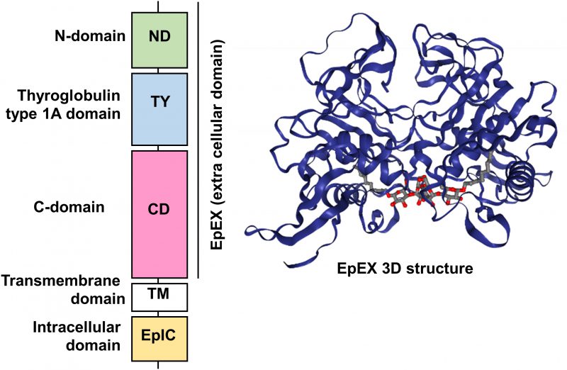
FIGURE 1: Schematic diagram of the domain structure of full length EpCAM protein and crystal structure of an extracellular EpCAM cis-homodimer according to Pavsic et al. Full length EpCAM consists of a N-terminal signal peptide (SP) followed by three compactly folded extracellular domains (N-Domain (ND), Thyroglobulin type 1A domain (TY) C-Domain CD), a single spanning transmembrane domain (TM) and a c-terminal intracellular domain (EpIC). Two EpCAM subunits form a heart-shaped dimer on the cell surface [11]. Protein Data Base entry 4MZV.
11. Pavsic M, Guncar G, Djinovic-Carugo K, Lenarcic B (2014). Crystal structure and its bearing towards an understanding of key biological functions of EpCAM. Nat Commun 5(4764. doi: 10.1038/ncomms5764