Back to article: Making the final cut: pathogenic amyloid-β peptide generation by γ-secretase
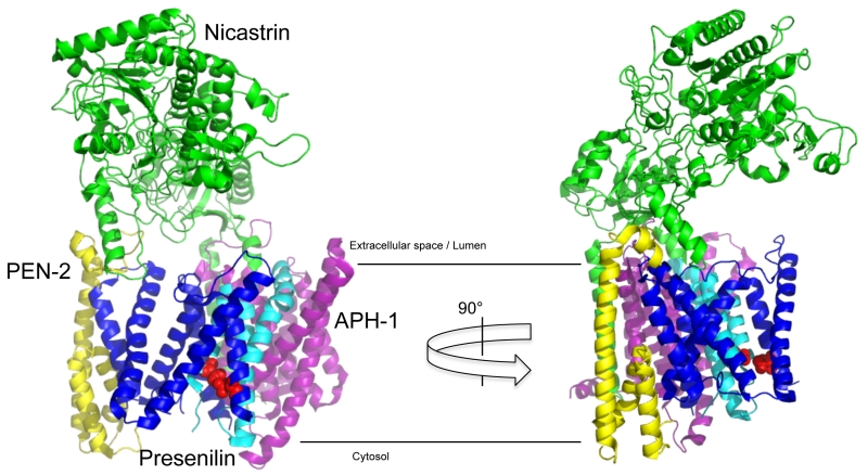
FIGURE 2: Structure of γ-secretase. The atomic resolution structure of γ-secretase (PDB: 5FN3) shows a membrane embedded core containing the catalytic subunit presenilin-1 cleaved into NTF (blue) and CTF (cyan) flanked by the subunits PEN-2 (yellow) and APH-1a (purple), which is covered by the large bilobar extracellular domain of the nicastrin subunit (green). This domain allows nicastrin to serve as gatekeeper controlling substrate access to the active site by excluding proteins with too large (or sterically incompatible) ectodomains. Red spheres depict the active sites aspartate residues.