Back to article: The role of hydrophobic matching on transmembrane helix packing in cells
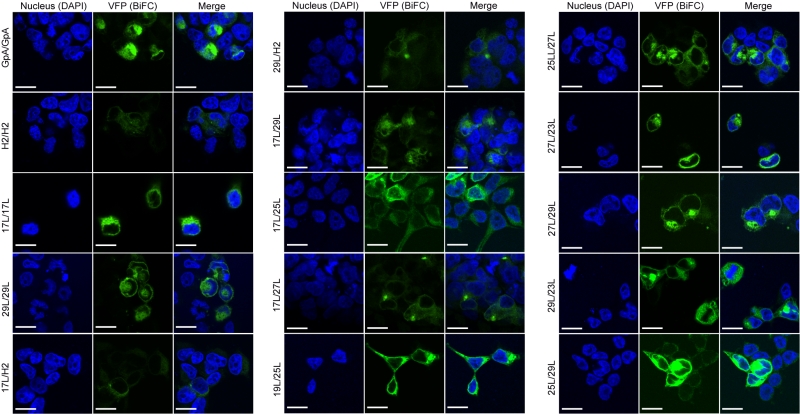
FIGURE 8: Confocal microscopy analysis for membrane dimer formation. Confocal microscopy of DAPI stained (blue) HEK293T cells expressing representative VN-VC combinations (GpA [VN-GpA/VC-GpA], 17L [VN-17L/VC-17L], 29L [VN-29L/VC-29L], 17L/H2 [VN-17L/VC-H2], 29L/H2 [VN-29L/VC-H2], 17L/29L [VN-17L/VC-29L]). Successful TM-driven oligomerization results in VFP reconstitution and fluorescent signal (green). Scale bar size is 16 µm.