Back to article: The role of hydrophobic matching on transmembrane helix packing in cells
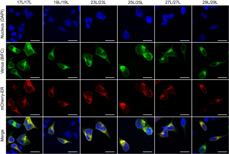
FIGURE 10: Homo-dimerization at the endoplasmic reticulum. Confocal microscopy of DAPI stained (blue) HEK293T cells expressing tested homo-dimers (17L [VN-17L/VC-17L], 19L [VN-19L/VC-19L], 23L [VN-23L/VC-23L], 25L [VN-25L/VC-25L], 27L [VN-27L/VC-27L], 29L [VN-29L/VC-29L]). TM-driven homo-oligomerization results in VFP reconstitution and fluorescent signal (green). Sec61α fused to mCherry fluorescent protein was used as ER marker (red). The presence of colocalization of red and green signals in the merge images was highlighted in yellow. Scale bar size was set to 16 µm.