Back to article: Dram1 regulates DNA damage-induced alternative autophagy
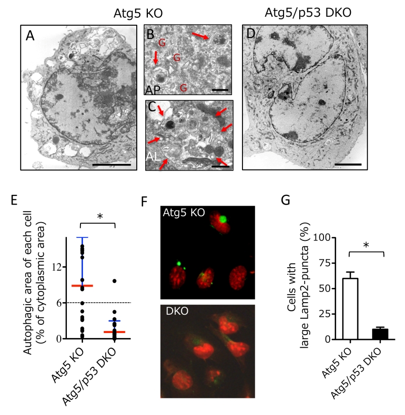
FIGURE 1: Induction of autophagy by etoposide in Atg5 KO MEFs, but not Atg5/p53 DKO MEFs. (A-D) Electron micrographs of Atg5 KO MEFs (A-C) and Atg5/p53 DKO MEFs (D) treated with etoposide (10 µM) for 18 h. In (A, D), bar = 5 µm. In (B), arrows indicate autophagosomes. “G” indicates Golgi membranes. Bar = 0.5 µm. In (C), the arrows are surrounding an autolysosome. Bar = 0.5 µm. (E) The percentage autophagic area of MEFs treated with etoposide. The indicated MEFs were treated with etoposide for 18 h and the autophagic area of each cell was calculated (n > 25 cells each). Red and blue lines indicate the mean and SD, respectively. The dotted line indicates the autophagic threshhold. *p < 0.05. (F) The indicated MEFs were treated with etoposide (10 µM) for 12 h. The cells were then examined for Lamp2 immunofluorescence (green) and nuclei were counterstained with propidium iodide (red). (G) The percentage of cells with large Lamp2 puncta is shown as the mean + SD (n = 4). *p < 0.05.