Back to article: Intravital imaging tumor screen used to identify novel metastasis-blocking therapeutic targets
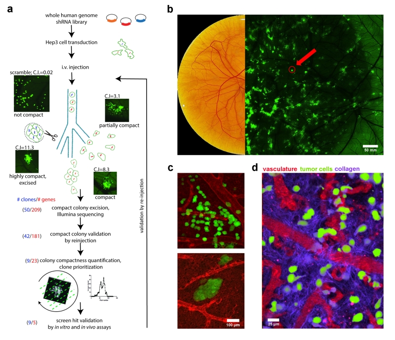
FIGURE 1: Chicken embryo as a platform for anti-metastatic target discovery. (a) Overview of intravital shRNA screen for drivers of human cancer metastasis. (b) Chicken CAM, fifteen days post fertilization. Left image shows brightfield view, right image shows a low-magnification, fluorescence image of GFP-expressing metastatic cancer cell colonies within the CAM tissue. Red arrow points to compact metastatic cancer cell colony. (c) High-resolution images of invasive (upper) and compact (lower) metastatic cancer cell colonies (green) and their associated vasculature (red) within the chicken CAM tissue. (d) Intravital imaging of cancer cells (green) and their interaction with vasculature (red) and the collagen matrix (purple) within the chicken CAM tissue. Collagen was visualized using second harmonic generation (SHG) imaging.