Back to article: Making the final cut: pathogenic amyloid-β peptide generation by γ-secretase
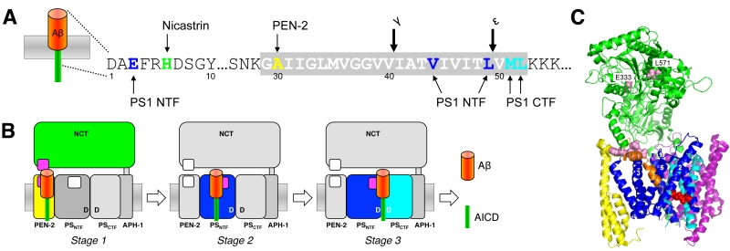
FIGURE 3: Substrate recruitment of C99 by γ-secretase. (A) Schematic representation of the most prominent C99 residues interacting with γ-secretase subunits as determined by site-directed photocrosslinking using the unnatural amino acid p-benzoyl-phenylalanine. (B) Model depicting the stepwise translocation of C99 from exosites (purple) in NCT and PEN-2 (stage 1) and the PS1 NTF (stage 2) to the active site (stage 3). (C) Structure of γ-secretase (5FN3) with a co-isolated α-helix (orange), which might represent a substrate-mimic. Red spheres depict the active site aspartate residues. Light pink spheres represent candidate sites for substrate interactions with NCT. Although not visible in this view, L571 is less buried than E333. Numbers indicate TMDs of the PS1 NTF that surround the substrate-mimicking α-helix.