Back to article: MiR-200c reprograms fibroblasts to recapitulate the phenotype of CAFs in breast cancer progression
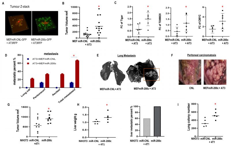
FIGURE 6: Effect of miR-200c overexpression in fibroblasts on primary breast cancer tumor growth and metastasis. (A-F) Co-injection of AT3-RFP cells with MEFs-GFP miR-200c or controls. Mice were sacrificed 6 weeks post-injection. (A) Tumor Z-stack images. (B) Tumor volume. (C) Immunoblot to assess the metabolic markers in the generated tumors. (D) Prevalence of metastatic disease. (E, F) Representative images for lung India ink staining and peritoneal metastasis are shown. (G-I) Co-injection of 4T1 cells and miR-200c NIH3T3 fibroblasts or controls. Mice were sacrificed 18 days post-co-injection. (G) Tumor volume. (H) Liver weight and metastatic percent. (I) Lung metastatic colonies. 6-TG resistant lung colonies quantified using image J.