Back to article: MiR-200c reprograms fibroblasts to recapitulate the phenotype of CAFs in breast cancer progression
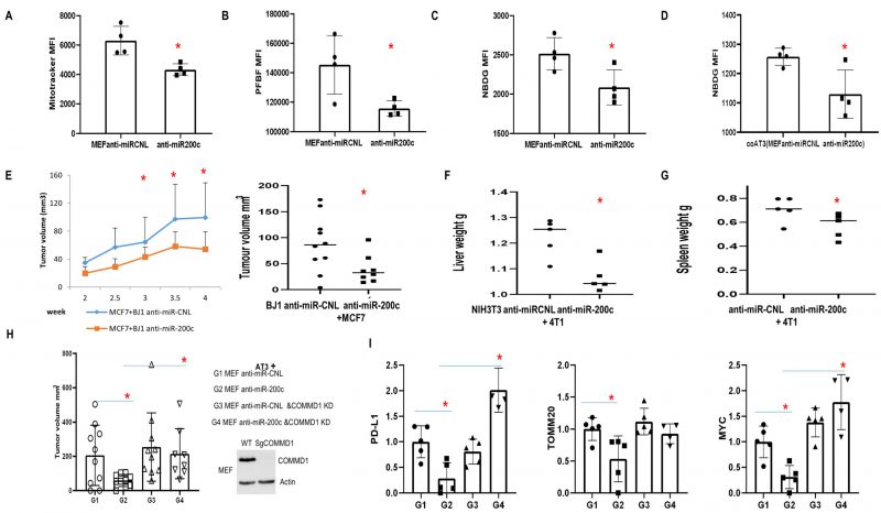
FIGURE 8: Effect of anti-miR-200c and COMMD1 KD in fibroblasts on primary breast cancer tumor growth and metastasis. (A-C) Flow data showing functional mitochondrial mass, PFBF and NBDG in MEF overexpressing anti-miR-200c versus control. (D) Flow data showing NBDG in AT3 cancer cells co-cultured with fibroblasts overexpressing anti-miR-200c versus control. (E) MCF7 cells co-injected with BJ1 anti-miR-200c or control. Tumor volume was measured at over time and at time of sacrifice (n=8-10, *p<0.05). (F, G) Liver and Spleen weight from mice co-injected with 4T1 and NIH 3T3 cells anti-miR-200c or control. (n=5-13, *p<0.05). (H, I) Co-injection of AT3 cells with MEFs with COMMD1 KD/WT overexpressing anti-miR-200c or controls. Mice were sacrificed 24 days post-injection. (H)Tumor volume. (n=8-10, *p<0.05). (I)Immunoblot to assess PD-L1, TOMM20 and MYC in the generated tumors.