Table of contents
Volume 2, Issue 2, pp. 20 - 39, February 2018
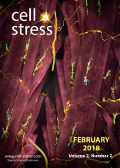
Cover: This month in
Cell Stress: The role of neuromuscular junctions in aging. Image depicts neuromuscular junctions in fruit fly muscles. Muscles are stained red, neurons and striated muscles are stained yellow. Credit: NICHD/Y.J. Kim and M. Serpe
CC BY 2.0 license. Image modified by
Cell Stress. The cover is published under the
CC BY 4.0 license.
Enlarge issue cover
Centriolar satellites prevent uncontrolled degradation of centrosome proteins: a speculative review
Nicolas Lecland and Andreas Merdes
Viewpoint |
page 20-24 | 10.15698/cst2018.02.122 | Full text | PDF |
Abstract
Centriolar satellites are small electron-dense structures in the cytoplasm, mostly surrounding the pericentriolar material. Initially viewed as shuttles for the transport of centrosomal proteins, they have been implicated in the assembly of the pericentriolar material and in ciliogenesis. Although numerous proteins have been identified as components of centriolar satellites, their molecular function remains unclear. In this review article, we discuss recent findings that characterize centriolar satellites as regulators of protein degradation pathways: by sequestering E3 ligase MIB1, deacetylase HDAC6, and proteins of the autophagy pathway, centriolar satellites may regulate the turnover of centrosomal and ciliary components, protecting them from removal via proteasomal degradation, autophagy, and aggresomes.
Rejuvenation of the aged neuromuscular junction by exercise
Tabita Kreko-Pierce and Benjamin A. Eaton
Reviews |
page 25-33 | 10.15698/cst2018.02.123 | Full text | PDF |
Abstract
Age-dependent declines in muscle function are observed across species. The loss of mobility resulting from the decline in muscle function represents an important health issue and a key determinant of quality of life for the elderly. It is believed that changes in the structure and function of the neuromuscular junction are important contributors to the observed declines in motor function with increased age. Numerous studies indicate that the aging muscle is an important contributor to the deterioration of the neuromuscular junction but the cellular and molecular mechanisms driving the degeneration of the synapse remain incompletely described. Importantly, growing data from both animal models and humans indicate that exercise can rejuvenate the neuromuscular junction and improve motor function. In this review we will focus on the role of muscle-derived neurotrophin signaling in the rejuvenation of the aged neuromuscular junction in response to exercise.
Salmonella Typhimurium infection: Type I Interferons integrate cellular networks to disintegrate macrophages
Nirmal Robinson
Microreviews |
page 37-39 | 10.15698/cst2018.02.125 | Full text | PDF |
Abstract
Type I interferons have immunomodulatory functions during infection with bacteria and viruses. They are vital for the host defense against viruses and extracellular bacteria. However, recent evidences show that IFN-I contributes to immunopathology during intracellular bacterial infection. We had previously shown that IFN-I receptor knock out mice (ifnar-/-) are less susceptible to S. Typhimurium infection and the macrophages are resistant to S. Typhimurium-induced cell death dependent on RIP kinases commonly known as necroptosis. We have now recently shown that IFN-I-signaling through the activation of RIP kinases and PGAM5 exacerbates necroptosis in Salmonella Typhimurium-infected macrophages by downregulating Nrf2-dependent cytoprotective response mechanisms [Hos et al, JCB 2017].
miR34a: a master regulator in the pathogenesis of bronchopulmonary dysplasia
Pragnya Das, Mansoor Ali Syed, Dilip Shah, Vineet Bhandari
Microreviews |
page 34-36 | 10.15698/cst2018.02.124 | Full text | PDF |
Abstract
Bronchopulmonary dysplasia (BPD) is the most common chronic lung disease in infants with lifelong pulmonary and neurodevelopmental consequences. The pathogenesis of BPD is contributed by genetic and environmental factors; among the latter, a critical contributor is exposure of the developing lung to hyperoxia. We have recently reported (Nat Comm 8:1173) that hyperoxia exposure in our in vitro and in vivo modeling systems of hyperoxia-induced lung injury (HALI) and BPD leads to an upregulation of the microRNA (miR) 34a. Utilizing genetic loss- and gain- of function strategies, we show that miR34a inhibition ameliorates the pulmonary phenotype of BPD (including BPD-associated pulmonary hypertension), at least in part, via one of the downstream targets of miR34a, namely Angiopoietin1/Tie 2 signaling. In addition, we demonstrate translational clinical significance of our findings by showing increased miR34a and decreased Ang1 expression in 3 independent cohorts of human lung samples.