Table of contents
Volume 2, Issue 9, pp. 219 - 241, September 2018
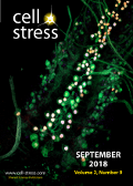
Cover: This month in
Cell Stress: The role of lamins in the maintenance of nervous system integrity. Image depicts insect larval central nervous system. Credit: NICHD/M. Sulkowski, licensed under a
CC BY 2.0 license. Image modified by
Cell Stress. The cover is published under the
CC BY 4.0 license.
Enlarge issue cover
Unweaving the role of nuclear Lamins in neural circuit integrity
Samantha L. Deal and Shinya Yamamoto
News and thoughts |
page 219-224 | 10.15698/cst2018.09.151 | Full text | PDF |
Abstract
Lamins are type-V intermediate filament proteins that comprise the nuclear lamina. Although once considered static structural components that provide physical support to the inner nuclear envelope, recent studies are revealing additional functional and regulatory roles for Lamins in chromatin organization, gene regulation, DNA repair, cell division and signal transduction. In this issue of Cell Stress, Oyston et al. (2018) reports the function of Lamin in the maintenance of nervous system integrity and neural circuit function using Drosophila. A number of laminopathies in humans exhibit age-dependent neurological phenotypes, but understanding how defects in specific neural cell types or circuitries contribute to patient phenotypes is very challenging. Drosophila provides a simple yet sophisticated system to begin untangling the vulnerability of diverse neuronal cell types and circuits against cellular stressors induced by defects in nuclear lamina organization.
Neuronal Lamin regulates motor circuit integrity and controls motor function and lifespan
Lisa J. Oyston, Yong Qi Lin, Thang M. Khuong, Qiao-Ping Wang, Man Tat Lau, Teleri Clark and G. Gregory Neely
Research Reports |
page 225-232 | 10.15698/cst2018.09.152 | Full text | PDF |
Abstract
Neuronal aging involves a progressive decline in cognitive abilities and loss of motor function. Mutations in human Lamin genes (LMNA, LMNB1, LMNB2) lead to a wide-range of diseases including muscular dystrophy, peripheral neuropathy and progeria. Here we investigate the role of neuronal Lamin in regulating age-related phenotypes. Neuronal targeting of Lamin led to shortened lifespan, progressive impairment of motor function and loss of dopaminergic (DA) neurons within the protocerebral anterior medial (PAM) cluster in the Drosophila melanogaster brain. Loss of neuronal Lamin caused an age-related decline in neural physiology, with slower neurotransmission and increased chance of motor circuit failure with age. Unexpectedly, Lamin-dependent decline in motor function was specific for the chemical synapses of the dorsal longitudinal muscle (DLM). Together these findings highlight a central role for Lamin dysfunction in regulating neuronal survival and motor circuit physiology during aging.
New insights into the role of soluble E-cadherin in tumor angiogenesis
Maggie K.S. Tang, Philip P. Ip and Alice S.T. Wong
Microreviews |
page 236-238 | 10.15698/cst2018.09.154 | Full text | PDF |
Abstract
A key to successful metastasis is the formation of new vasculature, known as angiogenesis. Therefore, it is of great interest to unravel the underlying molecular mechanisms of tumor angiogenesis. Cadherins are a major class of cell surface receptors. The loss of cadherins, especially E-cadherin, is a well-established marker for tumor metastasis. Loss of E-cadherin is also a defining characteristic of several carcinomas, such as lobular carcinoma of the breast, and de-differentiated endometrioid carcinoma of the endometrium and ovary, which are known to be associated with more aggressive tumor behavior. Although E-cadherin is synthesized as a transmembrane molecule, its extracellular domain can be enzymatically cleaved off and released as a soluble E-cadherin (sE-cad), and this accounts for the loss of E-cadherin function or expression that has been implicated in tumor progression and metastasis. Importantly, sE-cad is present at high levels in the serum and malignant ascites of ovarian carcinoma patients. Nevertheless, little is known about how this essential protein dictates metastasis. Hitherto, many studies have given attention only to the dominant negative role of the loss of E-cadherin in weakening cell-cell adhesion, however, it is not known if sE-cad has biological activity in itself. In addition, the release mechanism of sE-cad has remained elusive. Here we show for the first time that sE-cad is a pivotal regulator of angiogenesis. We further show that exosomes are a novel major platform for the cleavage and release of sE-cad in vitro, in vivo and in patients’ derived samples (Nat Commun, 9: 2270).
Red blood cell extracellular vesicles as robust carriers of RNA-based therapeutics
Chanh Tin Pham, Xin Zhang, Austin Lam, Minh TN Le
Microreviews |
page 239-241 | 10.15698/cst2018.09.155 | Full text | PDF |
Abstract
One of the major challenges of RNA-based therapeutics is the method for delivery of RNA molecules. In a recent article (Nat Commun 9(1):2359), we described a novel delivery platform for RNA-based drugs using red blood cell extracellular vesicles which can efficiently deliver both small and large RNAs to solid and liquid tumours. Our RBCEVs platform features exceptional merits over conventional RNA delivery methods in biosafety, biocompatibility, efficiency, accessibility, and cost effectiveness.
SKP2 and OTUD1 govern non-proteolytic ubiquitination of YAP that promotes YAP nuclear localization and activity
Fan Yao, Zhenna Xiao, Yutong Sun and Li Ma
Microreviews |
page 233-235 | 10.15698/cst2018.09.153 | Full text | PDF |
Abstract
Dysregulation of signaling pathways that control organ size, such as the AKT-mTOR and Hippo-YAP pathways, often leads to tumorigenesis and metastasis. The Hippo pathway effector YAP is a transcriptional co-activator overexpressed or activated in human tumors. Accumulating evidence has demonstrated that YAP promotes tumor initiation and/or progression in various types of cancer. YAP shuttles between the nucleus and the cytoplasm of the cell. When in the nucleus, YAP binds to transcription factors, such as SMAD, p73, RUNX, and the TEA domain (TEAD) family members, to activate gene transcription. The nuclear localization of YAP can be inhibited by the Hippo phosphorylation cascade and the cytoplasmic binding partners of YAP. In addition, YAP has previously been shown to be ubiquitinated by the SCFβ-TRCP complex and degraded by the proteasome. Recently, we discovered a novel mechanism by which non-proteolytic, K63-linked polyubiquitination of YAP promotes its nuclear localization, transcriptional activity, and growth-promoting function (Yao et al. Nat Commun 9:2269). Moreover, by screening ubiquitin E3 ligases implicated in K63-linked ubiquitination and a human deubiquitinase (DUB) library, we identified the SCFSKP2 complex and OTUD1, respectively, as the E3 ligase and the DUB that regulate this non-proteolytic ubiquitination without altering YAP protein level. Interestingly, this ubiquitination-mediated regulation of YAP is independent of Hippo pathway-mediated phosphorylation of YAP.