Back to article: Inflammation induced PD-L1-specific T cells
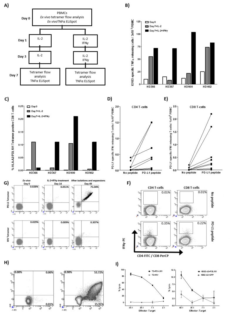
FIGURE 1: Pro-inflammatory cytokines induce expansion of PD-L1-specific T cells in vitro. (A) Experimental set up: PBMCs from four healthy donors were stimulated twice with IL-2 with or without IFN-γ. On day 7, cultures were examined for T-cell reactivity towards PD-L1 by either tetramer flow analysis or TNF-α-ELISPOT. (B) Tetramer analysis of PD-L1-specific CD8 T cells at day 0 (white bars) and at day 7 for cultures stimulated with IL-2 either without IFN-γ (black bars) or with IFN-γ (grey bars). Cells were stained with the tetramers HLA-A2/(PDL115-23; LLNAFTVTV)-PE/APC or HLA-A2/(HIV-1 pol476-484)-PE/APC. (C) TNF-α-ELISPOT responses against IO103 (PDL19-28; FMTYWHLLNAFTVTVPKDL) peptide at day 0 (white bars) and at day 7 for cultures stimulated with IL-2 either without IFN-γ (black bars) or with IFN-γ (grey bars). All experiments were performed in duplicates and the average number of IO103 induced spots (after subtraction of spots without added peptide) are calculated per 3×10⁵ PBMCs for each donor. (D, E) PBMCs from 8 healthy donors were stimulated three times with IL-2 and IFN-γ. On day 7, cells were incubated with and without PD-L1 peptides (PDL115-23 and IO103, PDL19-28) for 4 hr. IFN-γ-secreting PD-L1 peptide reactive CD4 and CD8 T-cells were analyzed using IFN-γ secretion assay. (F) Example of flow analysis of IFN-γ secreting PD-L1 specific CD4 and CD8 T cells from one healthy donor. (G) PBMCs from a healthy donors (HD400) were stimulated three times with IL-2 and IFN-γ. T cells were isolated three times using tetramers HLA-A2/ (PDL115-23; LLNAFTVTV)-PE and anti-PE MACS microbeads. Enriched T cells were expanded using high dose IL-2 (6,000 U/ml). On day 49, enriched T cells were analyzed for PD-L1 specificity by tetramer analysis, intracellular cytokine staining (ICS) and 51Cr-release cytotoxicity assay. Tetramer analysis of the resulting T-cell culture at day 0 (ex vivo), at day 14 (after three stimulations with IL-2 and IFN-γ), and at day 49 (after isolation and expansion) using the tetramers HLA-A2/(PDL115-23; LLNAFTVTV)-PE/APC, HLA-A2/(HIV-1 pol476-484)-PE. (H)On day 49, the resulting T-cell cultures were stimulated for 5 hours either with an irrelevant HIV peptide (HIV-1 pol476-484) or PD-L101 peptide (PDL115-23; LLNAFTVTV) before being analyzed for intracellular IFN-γ/TNF-α staining. (I) The resulting T-cell cultures were analyzed on day 49 using 51Cr-release cytotoxicity assay. Lysis of TAP-deficient T2 cells (pulsed with PD-L101 (PDL115-23)) or with irrelevant HIV peptide (HIV-1 pol476-484)) (left) or the HLA-A2-transfected leukemic cell line, K562, either transduced with the PD-L1 protein or with GFP (right).