Back to article: Exocytotic fusion pore under stress
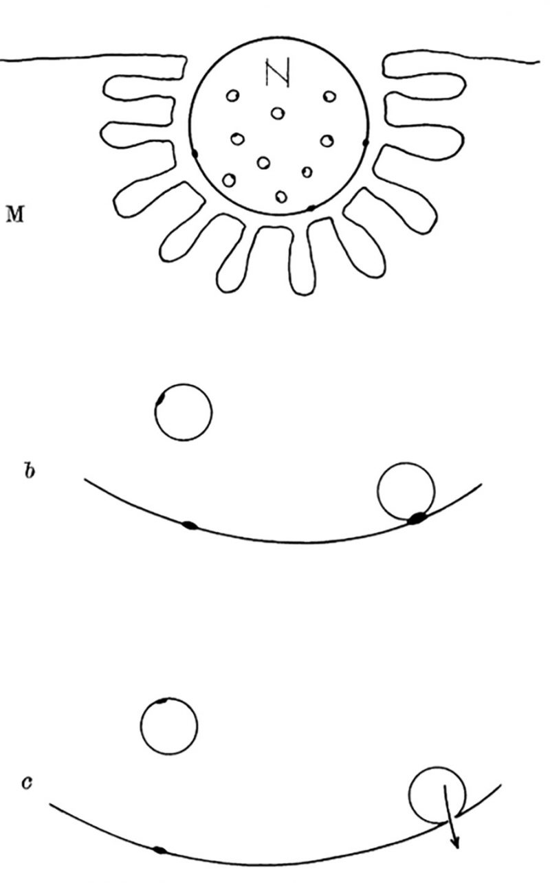
FIGURE 2: Diagram explaining quantal secretion of neurotransmitters by exocytosis of synaptic vesicles. Reaction molecules (fusion proteins) are indicated by black dots on vesicular and nerve membranes; a nerve impulse greatly increases the number of reactive sites in the terminal membrane by allowing calcium ions to penetrate it. N, motor nerve terminal; M, end-plate region of a muscle fibre. This scheme was presented by J. del Castillo and B. Katz at a Symposium at Gif-sur-Yvette in July 1955 [73].
73. Del Castillo J, Katz B (1957). La base “quantale” de la transmission neuomusculaire. In: Microphysiologie Comparee des Elements Excitables. Coll Int CNRS 67: 245-258.