Back to article: Targeting the TGFβ pathway in uterine carcinosarcoma
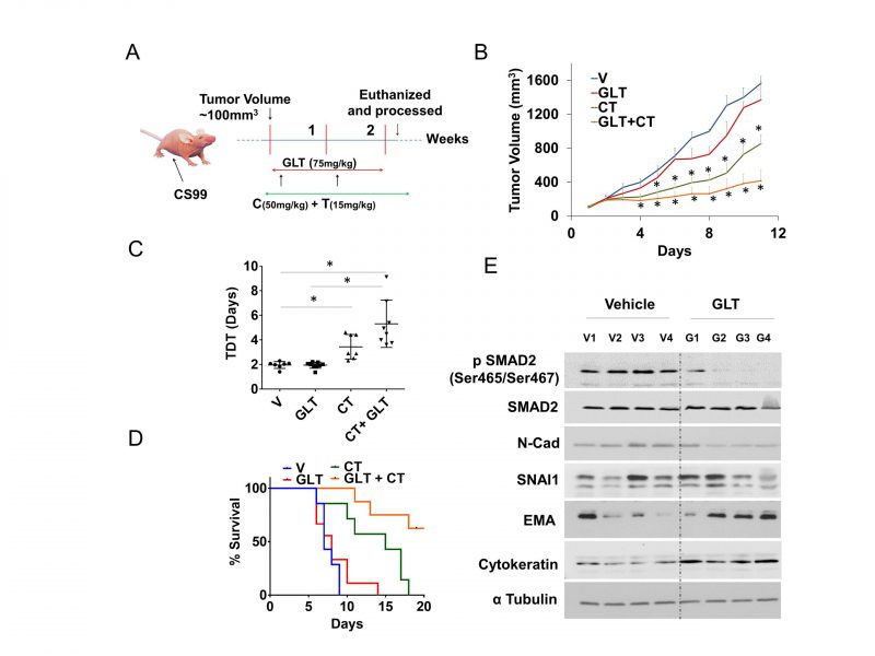
FIGURE 4: GLT sensitizes tumor cells to chemotherapy in a mouse model.(A) Female athymic nude mice were subcutaneously injected with CS-99 cells, once the tumor reached to ~100 mm3, tumor bearing mice were randomized into four groups (n = 7-10) and treated with either control vehicle (V), GLT, carboplatin & paclitaxel (CT), or GLT + CT. Treatment continued for two cycles (14 days), and mice were followed for survival. (B) Relative tumor volume normalized with V (vehicle) group. (C) Tumor doubling time (TDT) was calculated for each treatment groups according to Mehrara and colleagues [35]. Data are mean ± SD and *, P < 0.05 when comparing between indicated groups by one-way ANOVA (D) Percent survival was calculated by Kaplan–Meier method and P values determined by log-rank test based on the number of days the animals survived before the euthanization as per IACUC limits (humane endpoint). (E) Four tumor samples from vehicle treated control and GLT treated group were analyzed for the expression of TGFβ1 signaling and EMT markers using immunoblotting, alpha tubulin was used as loading control. Values are mean ±SD. *P<0.05.
35. Mehrara E, Forssell-Aronsson E, Ahlman H, Bernhardt P (2007). Specific growth rate versus doubling time for quantitative characterization of tumor growth rate. Cancer Res 67(8): 3970-3975. 10.1158/0008-5472.CAN-06-3822