Back to article: Fine intercellular connections in development: TNTs, cytonemes, or intercellular bridges?
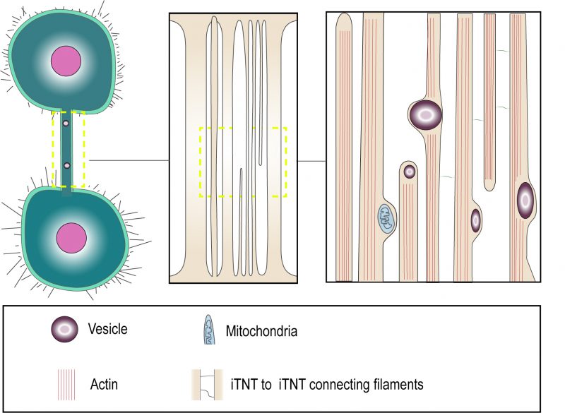
FIGURE 1: “Thick” and “thin” TNT connections. Cryo-electron microscopy shows that TNTs can either be a single thick connection or a bundle of thin individual TNTs (iTNTs). Both open-ended and closed-ended protrusions can be present within a bundle. Each iTNT contain actin bundles, can contain vesicles and mitochondria. Thin and short membrane threads connect several iTNTs, which appear to grow in opposite directions. Adapted from Sartori-Rupp et al. [31].
31. Sartori-Rupp A, Cordero Cervantes D, Pepe A, Delage E, Gousset K, Corroyer-Dulmont S, Schmitt C, Krijnse-Locker J, Zurzolo C (2019). Correlative cryo-electron microscopy reveals the structure of TNTs in neuronal cells. Nat Commun 10(1): 342. 10.1038/s41467-018-08178-7