Back to article: CRISPR-activation screen identified potassium channels for protection against mycotoxins through cell cycle progression and mitochondrial function
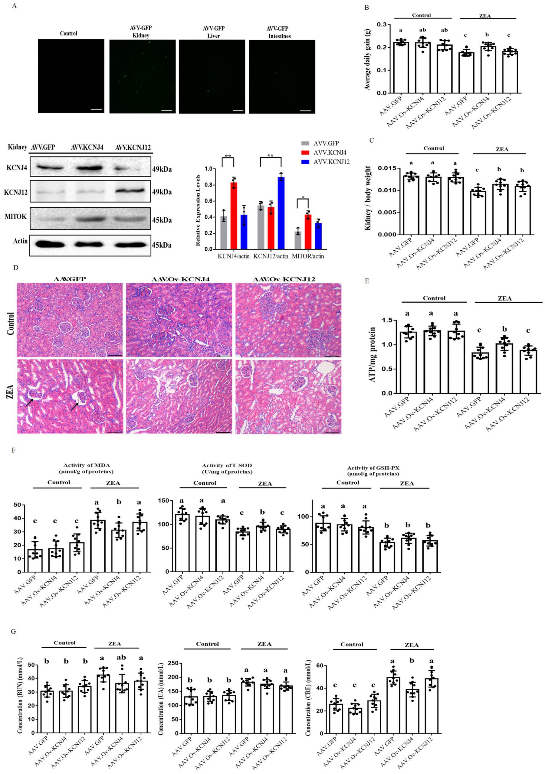
FIGURE 5: In vivo AAV manipulation of potassium channel proteins alleviated kidney damage and reduced activities of anti-oxidative enzymes. (A) Representative fluorescence photomicrographs of GFP expression in kidneys, live and intestines, and immunoblotting for KCNJ4, KCNJ12, MITOK and actin expression. (B) Change in the average daily gain. (C) Changes in the ratio of kidney weight to body weight in mice. (D) Detection of pathological changes in renal tissue by tissue section HE staining. (E) The levels of ATP of renal tissues of mice in each group were detected. (F) Oxidation and anti-oxidation parameters of renal tissues of mice in each group were detected by an oxidation kit. (G) The content of BUN, UA and CRE in the blood of mice. Animal data of each group are presented as the mean ± SEM (n=10). A two-way ANOVA with Bonferroni posttest was used for Figures B, C, E, F and G. A, B, C Values within a row with different superscripts differ significantly (P < 0.05); Other Data are presented as the mean ± SEM (n=3), calculated by Student t-test. Significant differences were designated as follows: p < 0.05 (*), p < 0.01 (**) and p < 0.001 (***).