Back to article: Striatal dual cholinergic /GABAergic transmission in Parkinson disease: friends or foes?
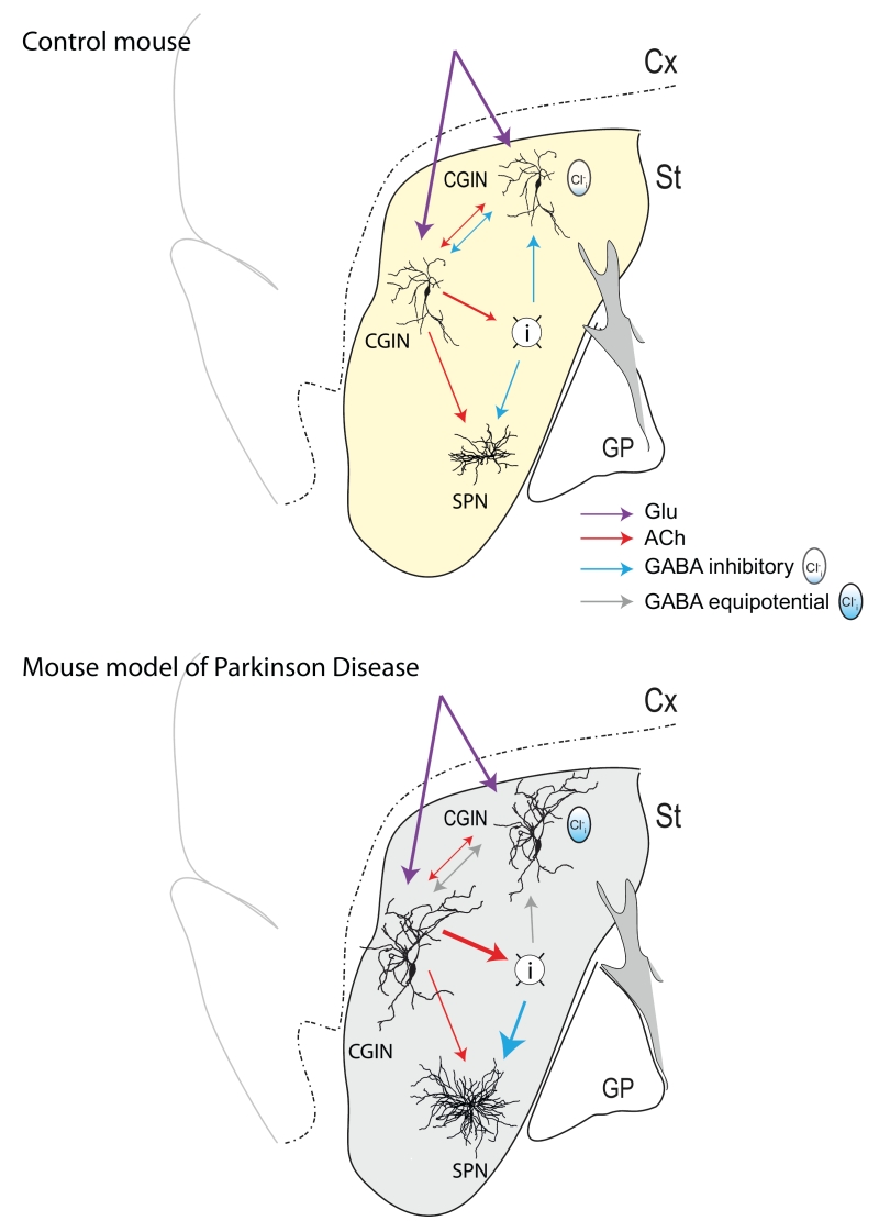
FIGURE 1: Schematic drawing of a mouse brain sagittal section showing sensory motor cortex (Cx), dorsal striatum (St), and Globus Pallidus (GP). Yellow indicates control dopaminergic innervation (top) whereas gray indicates depletion of dopaminergic terminals (bottom). Connections between CGINs, GABAergic Interneurons (I) and Spiny Projections Neurons (SPN) with different colors as indicated. Polarity of GABA actions depend on the intracellular chloride levels as illustrated (icon).