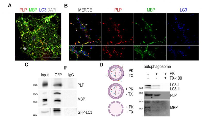
FIGURE 3: Myelin proteins PLP and MBP are detected in the autophagosomes. (A) Representative confocal image of a DIV2 primary OL, immunolabeled for MBP (green), PLP (red), LC3 (blue) and DAPI (grey). Rectangular boxes indicate areas magnified to the right. Scale bar: 30 μm. (B) Magnification images are of a single z-stack (0,5μm thick) and white arrows indicate the colocalized puncta in myelin membrane (top) and cytoplasmic processes (bottom). Scale bars: 2 μm. (C) Immunoprecipitation with antibodies against GFP in forebrain lysates from adult GFP-LC3 mice identifies MBP and PLP as interactors of LC3. (D) Shematic representation of the proteinase K protection assay and western blot analysis of isolated autophagosomes after proteinase K assay. Triton X-100 (TX-100) is used as a negative control. Autophagic marker LC3-II was protected from PK digestion, unless TX-100 was present. Both PLP and MBP seem to be protected from PK treatment.