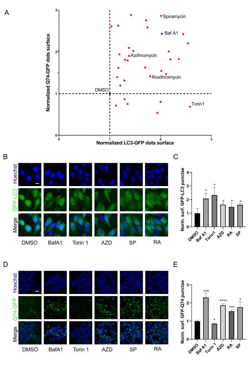
FIGURE 2: Identification of macrolide antibiotic as potent blockers of autophagic flux. (A) GFP-LC3B-expression U2OS cells and GFP-Q74-expression PC12 cells were treated with Torin 1 (300 nM), Baf A1 (100 nM) or macrolides (40 µM) for 6 h. (B, C) GFP-LC3-expression U2OS cells or (D, E) GFP-Q74-expression PC12 cells were treated with torin 1, Baf A1, AZD (40 µM), RA (40 µM) or SP (40 µM) for 6 h. Representative images are presented (B, D). The (C) GFP-LC3B or (E) GFP-Q74 puncta were assessed. Scale bars equal 10 μm. *P<0.05; **P < 0.01; ***P < 0.001; **** P < 0.0001 compared with DMSO/control.

