Table of contents
Volume 4, Issue 6, pp. 114 - 153, June 2020
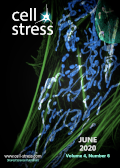
Cover: This month in
Cell Stress: Mitochondria in cancer. Image shows an osteosarcoma cell with mitochondria stained in blue. Public domain image by Dylan Burnette and Jennifer Lippincott-Schwartz, Eunice Kennedy Shriver National Institute of Child Health and Human Development, NIH. Image modified by
Cell Stress. The cover is published under the
CC BY 4.0 license.
Enlarge issue cover
Mitochondria in cancer
Debora Grasso, Luca X. Zampieri, Tânia Capelôa, Justine A. Van de Velde and Pierre Sonveaux
Reviews |
page 114-146 | 10.15698/cst2020.06.221 | Full text | PDF |
Abstract
The rediscovery and reinterpretation of the Warburg effect in the year 2000 occulted for almost a decade the key functions exerted by mitochondria in cancer cells. Until recent times, the scientific community indeed focused on constitutive glycolysis as a hallmark of cancer cells, which it is not, largely ignoring the contribution of mitochondria to the malignancy of oxidative and glycolytic cancer cells, being Warburgian or merely adapted to hypoxia. In this review, we highlight that mitochondria are not only powerhouses in some cancer cells, but also dynamic regulators of life, death, proliferation, motion and stemness in other types of cancer cells. Similar to the cells that host them, mitochondria are capable to adapt to tumoral conditions, and probably to evolve to ‘oncogenic mitochondria’ capable of transferring malignant capacities to recipient cells. In the wider quest of metabolic modulators of cancer, treatments have already been identified targeting mitochondria in cancer cells, but the field is still in infancy.
Mitochondria, mitophagy, and metabolic disease: towards assembling the puzzle
Zhiyong Chen, Marine Berquez and Alessandro Luciani
Microreviews |
page 147-150 | 10.15698/cst2020.06.222 | Full text | PDF |
Abstract
Dysregulation of the mitochondrial network in terminally differentiated cells contributes to a broad spectrum of disorders. Methylmalonic acidemia (MMA) is an autosomal recessive inborn error of intermediary metabolism caused by the deficiency of methylmalonyl-CoA mutase (MMUT) — a mitochondrial enzyme that mediates the degradation of certain amino acids and lipids. The loss of MMUT activity triggers an accumulation of toxic endogenous metabolites causing severe organ dysfunctions and life-threatening complications. How MMUT deficiency instigates mitochondrial distress and tissue damage remains poorly understood. Using cell and animal-based models, we recently discovered that MMUT deficiency disables the PINK1-induced translocation of PRKN/Parkin to MMA-damaged mitochondria, impeding their delivery and subsequent dismantling by macroautophagy/autophagy-lysosome degradation systems (Luciani et al. Nat Commun. 11(1):970). This promotes an accumulation of damaged and/or dysfunctional mitochondria that spark epithelial distress and tissue damage. Using a systems biology approach based on drug-disease network perturbation modeling, we predicted targetable pathways, whose modulation repairs mitochondrial dysfunctions in patient-derived kidney cells and ameliorates disease-relevant phenotypes in mmut-deficient zebrafish. These results unveil a link between primary MMUT deficiency, defective mitophagy, and cell distress, offering promising therapeutic avenues for MMA and other mitochondria-related diseases.
Towards understanding the role of Receptor Expression Enhancing Protein 5 (REEP5) in cardiac muscle and beyond
Shin-Haw Lee, Sina Hadipour-Lakmehsari and Anthony O. Gramolini
Microreviews |
page 151-153 | 10.15698/cst2020.06.223 | Full text | PDF |
Abstract
The sarco-endoplasmic reticulum (SR/ER) is the largest membrane-bound organelle in eukaryotic cells and plays important roles in essential cellular processes, and in development and progression of many cardiac diseases. However, many aspects of its structural organization remain largely unknown, particularly in cells with a highly differentiated SR/ER network. In a recently published study led by Lee et al. (Nat Commun 11(1):965), we reported a cardiac enriched SR/ER membrane protein REEP5 that is centrally involved in regulating SR/ER organization and cellular stress responses in cardiac myocytes. In vitro REEP5 depletion in mouse cardiac myocytes resulted in SR/ER membrane destabilization and luminal vacuolization along with decreased myocyte contractility and disrupted Ca2+ cycling. Further, in vivo CRISPR/Cas9-mediated REEP5 loss-of-function zebrafish mutants showed sensitized cardiac dysfunction to heart failure induction upon short-term verapamil treatment. Additionally, in vivo adeno-associated viral (AAV9)-induced REEP5 depletion in the mouse demonstrated cardiac dysfunction with dilated cardiac chambers, increased cardiac fibrosis, and reduced ejection fraction. These results demonstrate the critical role of REEP5 in SR/ER organization and function.